
HYPOTHALAMIC, PITUITARY AND OTHER SELLAR MASSES
Pituitary tumors , Pituitary adenomas :
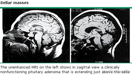
- Commonest tumors are pituitary adenomas. These are benign and their features depend on the cell type from which they occur.
- They may arise from PRL, GH, ACTH, TSH, LH and FSH producing cells r;esulting in features of hypersecretion of the hormones.
- There is autonomous hormone secretion which does not respond to the inhibitory feedback of these hormone levels.
- Many of these tumors do not produce hypersecretion features.
HYPOTHALAMIC, PITUITARY AND OTHER SELLAR MASSES
Causes of sellar masses
-
- Arachnoid
- Benign tumors
- Breast
- Carotid arteriovenous fistula
- Chordoma
- Craniopharyngioma
- Cysts
- Dermoid
- Germ cell tumor (ectopic pinealoma)
- Lactotroph hyperplasia (during pregnancy)
- Lung
- Lymphocytic hypophysitis
- Malignant tumors
- Meningiomas
- Metastatic
- Pituitary abscess
- Pituitary adenoma (most common sellar mass)
- Pituitary carcinoma (rare)
- Pituitary hyperplasia
- Primary
- Rathke’s cleft
- Sarcoma
- Somatotroph hyperplasia due to ectopic GHRH
- Thyrotroph and gonadotroph hyperplasia
Other neurologic symptoms —
- Cerebrospinal fluid rhinorrhea, caused by inferior extension of the adenoma, is an extremely uncommon presentation.
- Diplopia, induced by oculomotor nerve compression resulting from lateral extension of the adenoma.
- Headaches, presumably caused by expansion of the sella. The quality of the headache is not specific.
- Other neurologic symptoms that may cause a patient with a sellar mass to seek medical attention include:
- Parinaud syndrome, a constellation of neuroophthalmologic findings, most often paralysis of upward conjugate gaze, that result from ectopic pinealomas
- Pituitary apoplexy induced by sudden hemorrhage into the adenoma, causing excruciating headache and diplopia.
Pituitary carcinoma is rare. Craniopharyngiomas :
- These are derived from Rathke’s pouch. They arise near the pituitary stalk and extend to supracellar region.
- They are large, cystic and locally invasive.
- They may be calcified and seen on x-ray and CT. Age of presentation is less than 20 years.
- There is raised intracranial pressure with headache, vomiting, papilledema, hydrocephalus, visual field defects, cranial nerve damage, weight gain, personality changes, growth retardation.
- There may also be diabetes insipidus (if the posterior pituitary is involved).
Treatment of pituitary masses
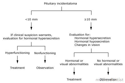
- Surgical resection and radiation after surgery. Patients may require lifelong pituitary hormone replacement.
Other tumors :
- Meningiomas, Histiocytosis X, Gliomas, Germinomas may also occur.
Lab diagnosis of tumors and masses
- · MRI, CT, visual field examination and other imaging techniques.
- · Histopathologic diagnosis of tumor after surgery.
- · Hormonal evaluation:
- 1. Basal PRL (prolactin)
- 2. IGF (insulin like growth factor)
- 3. 24-hour urinary free cortisol (UFC and or overnight oral dexamethasone 1 mg suppression test)
- 4. FSH and LH levels
- 5. Thyroid function tests.
Treatment of hypothalamic and pituitary masses
- Transsphenoidal surgery
- Stereotactic radiotherapy (gamma knife radiotherapy)
- Radiation
- Bromocriptine, the Dopamine agonist for hyperprolactinemia
- Estrogen replacement for bone loss and hypoestrogenem ia
- Growth hormone
- Thyroxine etc.