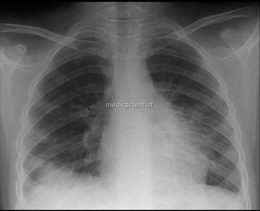
Radiographic x-ray Basic views (PA view) and (AP view)
Basic views–
- There are mainly two basic type of x-ray view ,
- Radiographic examination is the key to the diagnosis of many skeletal abnormalities,
- Obliteration of normal soft-tissue lines and the presence of a joint effusion are of particular importance,
- The radiographic findings are then correlated with the clinical history and the age and sex of the patient to arrive at a logical diagnosis,
- Posteroanterior view (PA view)
- Anteroposterior view (AP view)
1. Posteroanterior view (PA view)

Radiographic x-ray Basic views (PA view) and (AP view)
- The plain postero-anterior (PA) chest film is the most frequently requested radiological examination.
- Remember that the closer an object is to the film, the sharper are the borders.
- A current film is mandatory before proceeding to more complex investigations.
- Most of the important structures in the chest such as the heart and great vessels are located anteriorly.
- Comparison of the current film with old films is valuable and should always be undertaken if the old films are available.
- Therefore it is not surprising that the best way to take a chest radiograph is with the patient’s front against the film.
- The further away it is from the film, the more magnified and fuzzy is the shadow of the object (The Basics; experiment 2).
- Visualisation of the lungs is excellent because of the inherent contrast of the tissues of the thorax.
- Lateral films should not be undertaken routinely.
- The X-ray is shot from the patient’s back and is therefore
2. Anteroposterior view (AP view)
- Sometimes the patient is too sick to stand or sit for a PA view. so for emergency conditions,
- In this case, a lower quality AP view is taken.
- When patients are too sick to have an upright PA view of the chest, then an AP supine view is taken.
- A film is placed under the patient’s back and an X-ray is shot through the patient from the front.
- Recall that patients in this view are lying on their back. If an effusion is present, it will layer between the posterior chest wall and the lung due to gravity.
- Therefore, it appears larger than it really is and its borders are fuzzier, just like the finger in our experiment (The Basics; experiment 2),
- In this view, the heart is farther from the film.


