CLINICAL PRESENTATION
- Acute Myocardial Infarction is precipitated by exertion, exercise, emotional stress, medical and surgical disease and interventions.
- Circadian variation – AMI is more common in early morning hours due to increase in sympathetic tone and increased thrombolytic tendency from 4 to 12 AM.
- Acute myocardial infarction (AMI) is the rapid development of myocardial necrosis resulting from a sustained and complete absence of blood flow to a portion of the myocardium

Pain:
- Pain is the most common and typical symptom of AMI.
- There is very severe pain.
- It is located in central portion of chest and sometimes epigastrium.
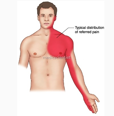
- It radiates to Arms; Abdomen; Jaw; Back and Neck.
- Pain may be present up to occiput or to umbilicus in front but not lower than umbilicus.
- It is a deep pain.
- It may be a severe discomfort, heaviness, squeezing, crushing pain.
Risk Factors
- Diabetes mellitus
- Dyslipidemia
- Advanced age
- Poorly controlled hypertension
- Tobacco use
- Family history of premature onset of coronary artery disease (CAD)
- Sedentary lifestyle
Chest pain is accompanied by :
- Weakness
- Sweating
- Pain may occur during rest.
- Painless MI – occurs in:
- Diabetes mellitus –
- old age
- Nausea
- Vomiting Anxiety
- Sense of doom.
- Pain occurs during exertion – is not relieved with rest.
- In Old age – Acute MI may present with acute aysponea (pulmonary edema).
Less Common presentations
- Loss of consciousness Confusion
- Extreme weakness Arrhythmia
- Pulmonary embolism Falling Blood pressure.
Associated Conditions
- Extracranial cerebrovascular disease
- Abdominal aortic aneurysm
- Atherosclerotic peripheral vascular disease
DIFFERENTIAL DIAGNOSIS
- Acute pericarditis Pulmonary embolism Acute aortic disease Costochond ritis Gastrointestinal disorders.
PHYSICAL FINDINGS
- Severe pain – Resulting in restless movements.
- Pallor
- Cold extremities Profuse sweating
- Severe substernal pain for >30 minutes
- In anterior infarction :
- Tachycardia
- Hypertension
- In Inferior infarction:
- Bradycardia
- Hypotension
Precordium –
- Apical impulse difficult to palpate.
- In Anterior wall M~ – dyskinetic bulge seen due to
- ‘nfarcted myocardium-in periapical area.
- S4 may be present S3 may be present
- Decreased intensity of heart sound Paradoxical splitting of 2nd heart sound
- Mid or late systolic murmur at apex due to MR due to dysfunction of Mitral valve apparatus. Pericardial friction rub.
- Decreased cardiac pulsations-decreased stroke volume.
- Temperature increased for a week.
- Feeble apex beat and increased JVP is Seen in right ventricular infarction.
- Systolic BP decreases by 10- 15 mmHg. –
ECG
- Acutephase
- Total occlusion results in ST elevation, with Q waves developing in hours or days.
- Q.waves ultimately develop.
- Q Wave MI is usually Transfmural infarction.
- In Sub total occlusion no ST elevation called Unstable angina. Non QMI is usually Non ST elevation MI.
- Non Q MI has positive serum cardiac marker with clinical Picture of MI.
- If no ST elevation is seen in a patient with clinical picture of MI with negative serum cardiac marker it is Unstable Angina.-
LAB FINDINGS
- 1.Acute few hrs to 7 days
- 2.Healing 7 to 28 days
- 3. Healed —- 29 days
- · ECG
- · Serum cardiac markers
- · Cardiac imaging
- · Non specific.
