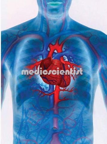
Now we are starting the series of – Cardiovascular System

The general appearance should be evaluated.
- <!–b1db3ec7868a4a669c04fa81b506a3b1–>
- A patient with low cardiac output looks tired and breathless.
- [intlink id=”225″ type=”post” /]
- A patient of pulmonary congestion / pulmonary edema is breathless and respiratory rate is increased.
- Central cyanosis with clubbing indicates right to left cardiac shunt.
- In heart failure the patient looks cyanosed with cool skin.
- A patient of infective endocarditis has petechiae, Osler’s nodes (Osler’s nodes are tender painful pulps of finger tips).
- The blood pressure is measured in both arms and with patient supine and upright.
- Heart rate is counted.
- All the peripheral pulses are examined like radial brachial, carotids, dorsalis pedis, posterior tibial, popliteal and femoral arteries.
- Fundoscopy should be done.
The abdomen is examined –
- abdominal aorta is palpated, pulsations of liver may be seen due to tricuspid regurgitation, epigastric pulsations may be seen due to right ventricular hypertrophy or abdominal aortic aneurysm.
- Ascites may be due to heart failure or constrictive pericarditis.
- systolic bruit over the kidneys may be due to renal artery stenosis and systemic hypertension.
- after this in next post we will discus deep examination of Cardiovascular System
This is the Beginning of Cardiac systems subscribe for all upcoming Posts of the Cardiovascular System
