Trypanosomiasis Chagas‘ disease Causes Diagnosis TREATMENT with Signs and Symptoms
African trypanosomiasis — African sleeping sickness, caused by Trypanosoma gambiense, and American trypanosomiasis —A disease caused by Trypanosoma cruzi and transmitted by the biting reduviid bug.
- The protozoan Trypanosoma is the cause of Chagas’ disease and sleeping sickness.
- American trypanosomiasis or Chagas‘ disease is caused by T. cruzi.
- Human African Trypanosomiasis or Sleeping Sickness is caused by T. brucei gambiense and T. brucei rhodesiense.
 |
| Trypanosomiasis Chagas‘ disease Causes Diagnosis |
CHAGAS’ DISEASE Trypanosomiasis
- Occurs in Americans. It caused by T. cruzi. A mild illness with fever occurs and subsides.
- Later in life, cardiac and gastrointestinal lesions develop and can even result in death.
- The typical feature of Chagas’ disease is that the acute illness subsides spontaneously and then may recur after an indefinite period.
 |
| Trypanosomiasis Chagas‘ disease Causative insect reduviid bugs fly |
Life cycle Trypanosomiasis
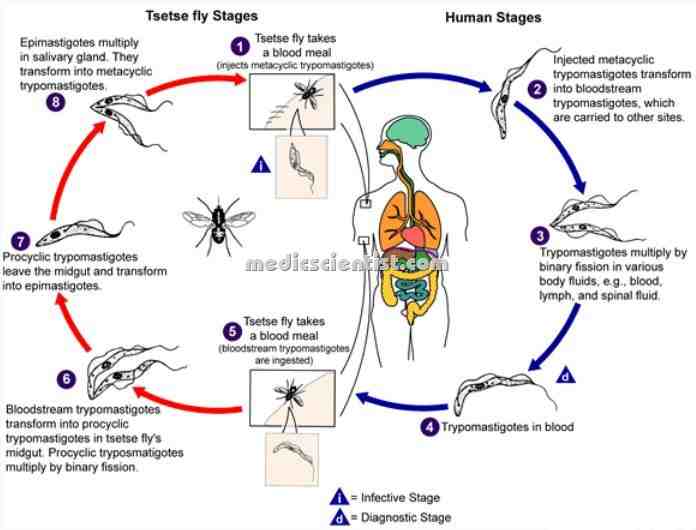
- Insects called reduviid bugs become infected by sucking blood from a human.
- The parasite multiplies, and is discharged with the feces of the insect, which can then contaminate skin, mucus membrane, and conjunctiva of a person.
- The parasite then enters through a break in the skin, mucus membrane and again multiplies.
 |
| Trypanosomiasis Chagas’ disease life cycle |
Pathology of Trypanosomiasis
- A chagoma is formed at the site of entry of the parasite. There is inflammation at the site.
- The organism disseminates via blood vessels and lymphatics and multiplies to form pseudocysts.
- The heart is affected, with destruction of heart valves and involvement of conduction system.
- The GIT is involved and there may be megacolon, involvement of the oesophagus and other parts of GIT.
 |
| Trypanosomiasis Chagas’ disease Pathology |
Clinical features of Trypanosomiasis
- · At first there is acute Chagas’ disease which subsides and many years later chronic Chagas’ disease appears.
- After one week of invasion by parasites, acute Chagas’ disease develops.
- · At the site of entry, there is erythema, swelling (Chagoma), and local lymphadenopathy.
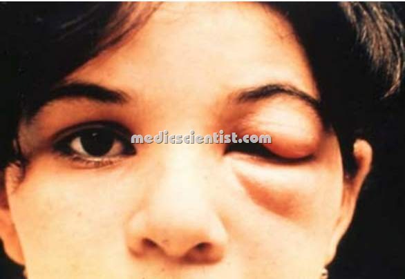
- · Romana‘s sign is classic finding of Chagas’ disease – there is unilateral painless edema around the eyes and is seen in conjunctival trypanosomiasis.
- There is malaise, fever, anorexia, edema over face and limbs. Rashes may also appear.
- · Generalized lymphadenopathy, hepatomegaly may occur.
- · Involvement of heart – Myocarditis, heart failure, conduction defects are seen.
- · In chronic Chagas’ disease there is dilated cardiomyopathy, arrhythmias like ventricular ectopics, thromboembolism, RBBB(right bundle branch block), AV block(atiroventricular heart block) heart failure.
- · Embolization from a mural thrombus in the heart, may occur.
- · Neurologic involvement – meningoencephalitis may occur.
- · GIT involvement – Dysphagia, odynophagia, chest pain, regurgitation, aspiration due to severe oesophageal dysfunction and aspiration pneumonitis, can occur.
- · There is weight loss, cachexia(wasting)
- · Death may occur due to septicemia, pulmonary infection, arrhythmias.
Diagnosis of Trypanosomiasis
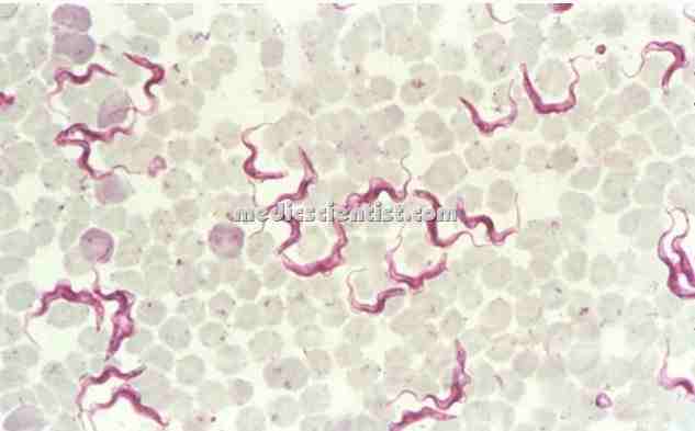
- · Fresh blood or buffy coat may show motile parasites in thin and thick smears. Acridine orange or Giemsa stain can be used.
- · PCR or hemoculture can be performed to diagnose Chagas’ disease.
- · Chronic Chagas’ disease is diagnosed by detection of specific antibodies for T cruzi.
- · Radiolabelled T cruzi antigens and electrophoresis are also used for diagnosis.
Trypanosomiasis Treatment
- Two drugs are available to treat Chagas’ disease. Nifurtimox and Benznidazole.
- They are very toxic and can cause nausea, vomiting, restlessness, seizures, polyneuritis.
- Dose of Nifurtimox is 10 mg/kg in 4 divided doses for 120 days.
- Cardiac transplantation is the only treatment for end-stage cardiomyopathy.
