Ischemic Heart Disease Physical Examination Lab Examination and Family history of premature IHD
Unstable Angina
- Unstable angina is angina pectoris with typical character of pain but certain atypical features like:
- It occurs at rest and lasts more than 10 minutes .2/. It is of new onset within the past 6 wks.
- It is more severe, prolonged and more frequent with a crescendo pattern.
Ischemic Cardiomyopathy Ischemic Heart Disease
- Ischemic cardiomyopathy is cardiomegaly and heart failure secondary to ischemic heart disease, ie is~hemic damage of left ventricular myocardium withCiLifany symptoms of ischemia.
Microvascular Angina Ischemic Heart Disease
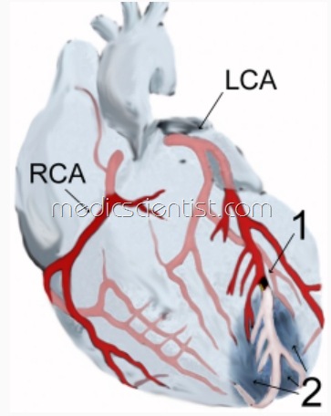
- Epicardial coronary arteries are conductance vessels. The main conductance vessels are the left main which divides into the left anterior descending and the left circumflex arteries, and the right coronary artery.
- Intramyocardial arterioles are resistance vessels.
- Abnormal constriction of resistance vessels results in microvascular angina.
Family history of premature IHD
- is defined as IHO at less than 45 years in male relative (blood relation) and less than 55 years in female relative.
- History of diabetes, hyperlipidemia, hypertension, cigarette smoking is significant.
Physical Examination Ischemic Heart Disease
- Often there are no findings.
- There may be – xanthomas (hypo-pigmented scars on medial side of upper eyelid-s), diabetic skin lesions, anaemia,-thyroid diseases, signs of ggarette smok-
- !ng ~ dark lips and fingers
- Thickened arteries
- Absent or feeble pulses Cardiomegaly
- LV Akinesia or Dyskinesia
- Fundus examination – AV nicking, increased light reflex, haemorrhages and exudates
- arterial bruits suggestive of atherosclerosis
Physical findings at the time of angina
- Transient LVF
- S3′ S4′
- Apical dyskinesia
- MR
- Pulmonary edema.
Lab Examination Ischemic Heart Disease
- Urine – for Diabetes and Renal disease Lipid profile – TC, TG, LDL, HDL
- Glucose, Creatinine and Hematocrit Thyroid function.
Chest X-ray –
- LVH, LVF, Pulmonary edema, Ventricular aneurysm.
ECG –
- shows -changes in only 50% of angina patients.
- ST-T – changes suggestive of angina are common to many diseases.
- Changes related to angina may appear only at the time of angina.
- ST segment is depressed in angina but elevated in MI and Prinzmetal angina.


