Aortic Regurgitation SYMPTOMS of Aortic Regurgitation Aortic Regurgitation PHYSICAL FINDINGS Aortic Regurgitation TREATMENT
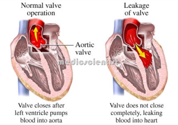
Aortic Regurgitation or aortic incompetence is a regurgitation of blood into the left ventricle during diastole due to defective closure of the aortic valve. A backward flowing, as in the return of solids or fluids to the mouth from the stomach or the backflow of blood through a defective heart valve. The long-term prognosis of patients with chronic aortic regurgitation (AR), particularly the favourable outlook of those who are asymptomatic, is well documented. Regurgitation caused not by valvular disorder but by dilatation of ventricles, the great vessels, or valve rings. AR is more predominant in males.
AR with mitral valve disease is more common in females.

Aortic Regurgitation ETIOLOGY
- Rheumatic heart disease
- Infective endocarditis especially over bicuspid aortic valve, and rheumatic heart disease
- Congenital bicuspid aortic valve with calcification, infective endocarditis or fibrosis leading to AR
- VSD with prolapse of aortic cusps
- Rheumatoid spondylitis
- Traumatic AR especially non penetrating injuries Cystic medial necrosis as in Marfan syndrome Osteogenesis imperfecta Syphilis
- Congenital subaortic stenosis with AR
- AR with AS is usually found in rheumatic aortic valve = 3Case,
- In Syphilitic AR there is no AS but narrowin of coronary ostia leading to coronary ischemia ,

Aortic Regurgitation PATHO PHYSIOLOGY
- – ventricular diastole, a part of the stroke volume ejected into the aorta regurgitates back into the LV, volume of blood in LV increases leading to dilatation of LV. The dilatation and hypertrophy of LV increasing the stroke volume and the AR is sated.
- As the time passes, the LV end diastolic volume increases, Diastolic pressure increases, ejection fraction
- these LV hypertrophy as well as dilatation resulting in a large heart. There is progressive LV dilatation leading tp left ventricular failure.
SYMPTOMS of Aortic Regurgitation
- There may be history suggestive of etiology e.g.
- Spondylitis, rheumatic fever syphilis etc.
- Palpitations – due to forceful apex, sinus tachycardia, VPCs, hyperdynamic circulation.
- Dyspnoea – exertional dyspnoea, orthopnea, paroxysmal nocturnal dyspnoea, dyspnoea at rest.
- Angina – due to LVH LV dilatation, coronar osteal narrowin , decreased diastolic filling of coronary arteries .
- Diaphoresis (Increased sweating) .
- Systemic venous congestion – hepatomegaly and ankle edema .
Aortic Regurgitation PHYSICAL FINDINGS
- There may be nodding movements of the head.
- Visible pulsations of large arteries in the neck.
- Features of Marfan syndrome may be present.
Arterial Pulse –
- There is a water-hammer collapsing pulse, also called Corrigan‘s pulse. This is a ig vo ume pulse with a rapid rise and a rapid fall.
- An alternate redness and paling of the nails may be seen on applying pressure to the tip of the nail called Quincke‘s pulse.
- Pistol-shot sounds can be heard over the femoral arteries called Traube‘s sign.
- There is a to-and-fro murmur over the femoral artery when compressed with a stethoscope called Duroziez murmur.
Blood pressure –
- The systolic pressure is high and the diastolic pressure is low, sometimes as low as 0 mmHg.
- However the level at which there is muffling of Korotkoff sounds is taken as the diastolic blood pressure.
- The wider the ulse ressure the more severe is theAR till the LV fail.
- When there is LV failure the LV end diastolic pressure rises and the diastolic blood pressure begins to increase.
Palpation of precordium –
- The LV apical impulse is shifted laterally and inferiorly.
- The apex impulse is hyperdynamic i.e. occupying an area more than 2.5 cm, ill-sustained and forceful.
- There is often a systolic thrill at the base of the heart and along the carotids in the neck.
- The carotid arterial pulse has two systolic waves called bisferiens pulse in severe AR, and also in AR with AS.
Auscultation
- The A2 or aortic closure sound is soft or absent.
- 53 or systolic ejection sound is present (due to poor LV compliance).
- 54 may also be audible.
Diastolic Murmur:
- Murmur of AR is high-pitched, blowing, decrescendo diastolic murmur heard best in left third intercostal space near the sternum and radiating downwards.
- The murmur is heard in early diastole but may be holodiastolic and loud in severe AR.
- The murmur is etter heard with the diaphragm of stethoscope, with the patient sitting up and leaning forwards, and with breath held in expiration.
- The murmur of AR is heard better on the left edge of sternum in valvular AR.
- The murmur of AR is heard better on the right side of sternum in AR due to aortic root disease.
- The murmur may be cooing or musical if there is rupture or perforation of cusp.
Systolic Murmur:
- A midsystolic ejection murmur is common in AR.
- It radiates along the carotids and is a functional murmur of relative aortic stenosis.
- It is higher pitched, less rasping than in organic AS.
- In functional AS, there is no accompanying reverse or paradoxical split of 52′
Austin-Flint Murmur:
- A mid-diastolic murmur which may be soft, low-pitched, and not accompanied by a thrill, may be heard at the apex due to diastolic displacement of the anterior mitral valve leaflet by the regurgitant stream. It is called Austin-Flint murmur.
- This murmur is not due to mitral valve obstruction.
- There is no opening snap or loud 51 or diastolic thrill as in MS.
Aortic Regurgitation INVESTIGATIONS

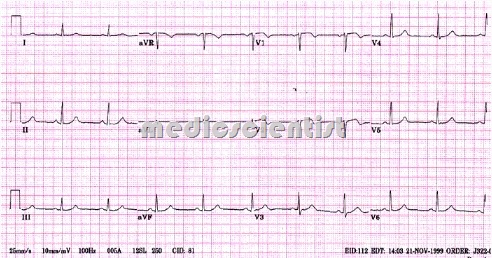
ECG –
- LVH, ST depression, T inversion, or tall T waves of diastolic overloadl and left axis deviation may be seen.

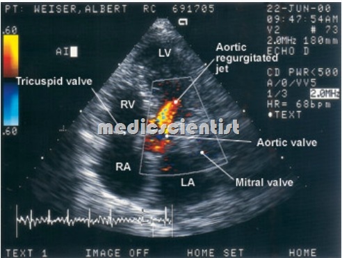
Echocardiogram –
- Rapid, high frequency fluttering of anterior mitral leaflet due to regurgitant jet is seen.
- Disease of the aortic valve apparatus or aortic root is seen.
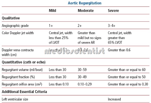
- Severity of AR can be assessed by Doppler.

X-ray –
- LVH is seen in the frontal view as well as in LAO and lateral views. It can be seen encroaching on the spine.
Catheterization and angiography –
- is always done before surgery to evaluate coronary disease and other associated abnormalities.
Aortic Regurgitation TREATMENT
- For heart failure – digitalis glycosides, salt restriction, diuretics, vasodilators like ACE inhibitors are given.
- Vasodilators and digitalis may be given even without LV failure in severe AR.
- Cardiac arrhythmias and infection must be treated promptly .
- Nitroglycerine, long-acting nitrates and long acting nifedipine relieve symptoms and delay surgery.
- Regular echocardiographic evaluation should be done at least every 6 months.
- Operation is advised when there are symptoms or LV dysfunction develops.
Indications for operation
- is LV EF (left ventricular ejection fraction) <55% or LV end systolic volume more than 55 ml / m2•
- Tissue prosthesis or mechanical valves may be implanted.
- When mechanical prosthetic valves are implanted, then regular evaluation of prothrombin time must be done and anticoagulants given lifelong.
