Examination of arterial pressure pulse

- The arterial pulse has an anacrotic shoulder on the ascending limb and then a rounded peak, then a downward deflection called incisura and then the pulse wave gradually comes down.
- In peripheral pulses the anacrotic shoulder is not seen and incisura is replaced by dicrotic notch which is smooth.
- [intlink id=”546″ type=”post” /]
- [intlink id=”225″ type=”post” /]
The different types of pulses are:
Pulsus parvus –
small, weak pulse due to decreased stroke volume and narrow pulse pressure.
Pulsus tardus
in aortic stenosis is a slow pulse with a delayed peak.
Hyperkinetic pulse –
- due to increased LV stroke volume, wide pulse pressure as in CHB, or hyperkinetic circulation like anaemia, exercise, fever, POA, AR, MR, and VSO.
Bisferiens pulse
- has two systolic peaks, as in AR and hypertrophic cardiomyopathy.
Pulsus alternans
- is a pulse with alternate tall and short peaks due to left ventricular failure.
Pulsus bigeminus
- is due to premature ventricular contraction after every beat, giving rise to a pulse coming in pairs.
Pulsus paradoxus –
- there is an exaggerated fall in systolic arterial pressure during inspiration. The fall is more than 10 mmHg so that the peripheral pulse may be seen to disappear during inspiration.
Radiofemoral delay–
- Simultaneous palpation of radial and femoral arterial pulses may show that the femoral pulse is delayed and weak. This is found in coarctation of aorta.

Examination of Jugular Venous Pulse (JVP)
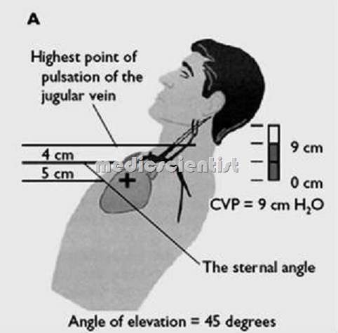
- The JVP is best seen in right internal jugular vein with the trunk inclined at 30 to 45°. With high JVP the trunk is elevated to 90° to see the upper level of venous pulse.
- The head is flexed a little to relax the neck veins and a tangential light from a torch is thrown across the skin over the vein to see the internal jugular vein.
- The JVP has 3 positive waves and 2 negative troughs.
- The positive waves are a, c, v and the negative waves are x and y.
- The ‘a’ wave is produced by right atrial contraction.
- The ‘c’ wave is produced by bulging of tricuspid valve into the right atrium during right ventricular systole and also due to the pulsation of carotid artery adjacent to jugular vein.
- The ‘x’ descent is due to atrial relaxation.
- The positive ‘v’ wave is due to filling of the right atrium when the tricuspid valve is closed.
- The ‘y’ descent is due to opening of tricuspid valve and emptying of the right atrium.
Causes of large ‘a’ wave
- Tricuspid stenosis
- Pulmonary hypertension
- Pulmonary stenosis
- Cannon ‘a’ waves (giant ‘a’ waves) in junctional rhythm and complete heart block.
Causes of absent ‘a’ wave
- Atrial fibrillation
Causes of prominent ‘x‘ descent
- Constrictive pericarditis
Causes of loss of ‘x’ descent
- Tricuspid regurgitation
Causes of ‘cv’ waves
- Triscuspid regurgitation (due to no ‘x’ descent)
Causes of deep ‘Y‘ descent
- Constrictive pericarditis
- Severe TR (tricuspid regurgitation)
- Severe RVF (right ventricular failure)
Causes of slow ‘Y‘ descent
- TS (tricuspid stenosis)
- Right atrial myxoma
Normal JVP (Jugular Venous Pressure)
- Normal JVP is measured from the sternal angle.
- The vertical distance between the top of the venous column and the level of sternal angle is usually less than 3 cm. This distance is 3 cm + 5 cm = 8 cm from the centre of right atrium.
- High venous pressure is seen in increased RV diastolic pressure.
Abdominojugular reflux test
- The palm of hand is placed over the abdomen and pressure is applied for 10 secs. When the right heart is failing, the upper level of venous pulse increases. An increase in NP and a rapid fall of 4 cm on release of pressure is found in right sided heart failure secondary to elevated left heart filling pressures.
Kussmaul’s sign
- An increase in JVP during inspiratio is called Kussmaul’s sign seen in
- constrictive pericarditis,
- RV infarction and RVF.
