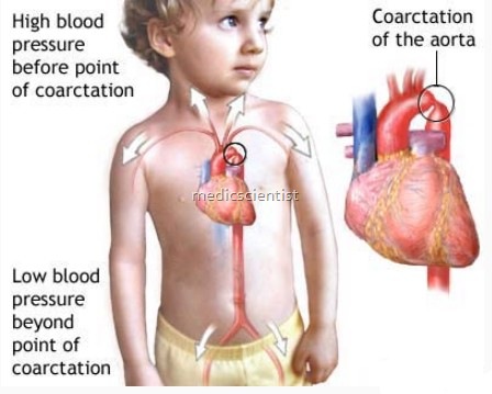
CYANOTIC CONGENITAL HEART DISEASE Coarctation of the Aorta Clinical features
coarctation of aorta means a narrowing of the aorta just distal to left subclavian artery, proximal (preductal) or distal (post ductal) to insertion of ductus arteriosus. A narrowing (discrete or of varying lengths) of the aorta at any point but usually located just distal to the left subclavian artery at the junction of the ligamentum arteriosum; primary effects are cardiovascular.
- Patient is acyanotic.
- The blood pressure proximal to the narrowing is increased so that there is hypertension in upper extremities.
- The coarctation may rarely be between left common carotid and left subclavian artery so that blood pressure in right upper limb is more than left.
- Coarctation may be mild or severe or there may be even total obliteration of lumen.
- It may be diagnosed in infancy, adolescents or adults.
- Lowest long-term mortality rates are observed in patients having surgery between 1 and 5 years of age.
- Condition is noted more commonly in infants than in adults.
- A greater risk of complications is possible if correction is delayed beyond early childhood. Often the diagnosis itself is delayed.
- Even with correction, higher incidence of miscarriage and HTN occurs in pregnancy.
- Uncorrected coarctation or restenosis carries a high risk of aortic rupture or dissection and cerebral hemorrhage (ruptured aneurysm, circle of Willis) but a smaller risk of preeclampsia than with other forms of HTN.

Clinical features Coarctation of the Aorta
- Male : Female ratio is 5 : 1.
- Coarctation of aorta is more common in men.
- Abdominal coarctation is more common in women.
- Most patients are asymptomatic.
- There is systolic hypertension in the upper extremities.
- General condition may be satisfactory or patient may become acutely ill when the ductus constricts. The trunk and lower limbs are deprived of arterial supply as pulmonary arterial blood goes into the lungs and left side of the heart.
- A collateral circulation develops around the coarctation.
- also there are continuous murmurs heard throughout the chest
- There is intermittent claudication.
- Cold lower extremities may be present.
- The blood pressure proximal to the narrowing is increased so that there is hypertension in upper extremities.
- ‘The systolic blood ressure is higher by 20 mmHg in the upper limbs compared to theiower:-
- Classic finding of coarctation of aorta is a radiofemoral
- delay of pulse timing.
- There is marked dyspnoea.
- Lower limbs may appear cyanosed.
- There may also be narrowing of abdominal aorta involving renal arteries, leading to severehypertension.
- Lower extremities may be smaller than upper. Femoral pulses are weak or absent .
- When coarctation is proximal to left subclavian ar~ery the left upper limb pulses are absent.
Associated Conditionswith Coarctation of the Aorta
- Bicuspid aortic valve: 85%
- Patent ductus arteriosus: 65%
- Mitral valve abnormalities
- Aortic stenosis and insufficiency
- Ventricular septal defect: 30–35%
- Aneurysm of circle of Willis
- Neurofibromatosis I
- Acquired intercostal aneurysms
- Subvalvular aortic stenosis
- Transposition of great vessels or double outlet right ventricle
