Mitral Stenosis Treatment of Mitral Stenosis causes and Etiology of Mitral Stenosis with Symptoms of Mitral Stenosis
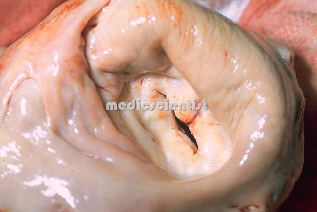
Mitral Stenosis is narrowing of the mitral valve due to which there is a restriction of blood flow from left atrium to left ventricle. It is more common in females. Mitral stenosis (MS), resulting from thickening and immobility of the mitral valve leaflets, In most adults, previous bouts of rheumatic carditis are responsible for the lesion. Mitral stenosis causes an obstruction in blood flow from the left atrium to left ventricle.
- The abnormality of the valve may predispose patients to infective endocarditis; to left atrial enlargement and atrial arrhythmias; or to left ventricular failure.
Mitral Stenosis Etiology of Mitral Stenosis Symptoms of Mitral Stenosis Treatment of Mitral Stenosis
ETIOLOGY of Mitral Stenosis
- In the great majority of cases, mitral stenosis has resulted from rheumatic involvement of the mitral valve
- Pure MS of rheumatic origin i.e. rheumatic heart disease is very common, and occurs in 40% of patients of RHD.
- Mitral stenosis with mitral regurgitation present together in a patient is usually of rheumatic origin.
- Congenital MS is rare.
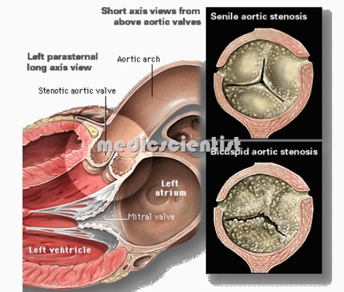
- Mitral stenosis with aortic valve disease is very common.
- The marked predominance of rheumatic carditis as a cause of MS in developed countries will not be so applicable in the future given the marked reduction in the incidence of rheumatic fever
- MS with AR is common in RHD. MS can also occur due to calcification of valve.
Functional MS Mitral Stenosis
- There is no organic disease of the mitral valve but there may be a relative stenosis due to increased volume of blood in left atrium as in MR,
- or due to inflammation of the valve as in rheumatic valvulitis(the mid-diastolic murmur is called Carey Coombs’ murmur)
Mitral Stenosis Etiology of Mitral Stenosis Symptoms of Mitral Stenosis Treatment of Mitral Stenosis
Features of Rheumatic mitral stenosis
- VaIve leaflets are thickened.
- There is fibrosis and calcification of different parts of valve apparatus.
- There is fusion of mitral commissures, and fusion of chordae tendinae with shortening.
- Valve cusps are rigid.
- There is narrowing of the valve opening.
- Immobility of leaflets leads to further narrowing.
Embolization in Mitral stenosis
- This is common due to :
- 1. Embolization of thrombus from enlarged left atrium
- 2. A piece of calcific valve embolizing to systemic circulation
- 3. Thrombus from LA embolizes when atrial fibrillation is converted to sinus rhythm.
- In mitral stenosis there is a stasis of blood in the left atrium and so a thrombus forms in left atrium especially in the left atrial appendage.
- Embolization of this thrombus occurs because the LA which was not contracting due to AF (atrial fibrillation), suddenly contracts when sinus rhythm is restored, resulting in breaking of a piece of thrombus and traveling through mitral valve and aorta to systemic circulation.
Mitral Stenosis Etiology of Mitral Stenosis Symptoms of Mitral Stenosis Treatment of Mitral Stenosis
Pathophysiology of Mitral Stenosis
- Normal mitral valve orifice is 4-6 cm2• When mitral valve is 1 cm2 or less, called critical MS the LA pressure and wedge pressure is 25 mmHg or more, resulting in dyspnoea, pulmonary edema and right ventricular failure.
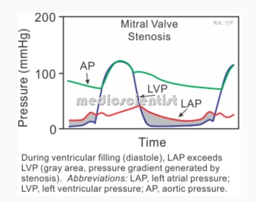
- The primary hemodynamic consequence of MS is a pressure gradient between the left atrium and left ventricle in diastole.
- Atrial fibrillation (AF) is common in patients with MS due to the elevation of left atrial pressure and consequent left atrial enlargement.
- With isolated MS, the left ventricular systolic and diastolic pressures are usually normal.
- Due to narrowing of the mitral valve, LA pressure rises and this is reflected via the pulmonary veins to the pulmonary vascular bed and pulmonary arteries, to the origin of pulmonary arteries and pulmonary trunk, to right ventricle. This results in increased LA pressure, and increased pulmonary wedge pressure.
- The pulmonary wedge pressure is the pressure of the pulmonary venous side and LA as recorded from the right side. This is done by a catheter inserted from a peripheral vein to the RA, RV and then pulmonary artery right up to the point where it cannot go any further.
- Hypertrophy of the pulmonary artery muscular layer Organic obliterating changes in the pulmonary vascular bed
- In MS the LV ejection fraction is normal and LV functions are normal.
- The pulmonary wedge pressure, pulmonary vascular resistance, pulmonary arterial pressure, RV end-diastolic pressure, RV volume are increased in MS.
Pulmonary Hypertension (PH)
- PH is a very important feature of MS. Pulmonary hypertension results from:
- Hypertrophy of the pulmonary artery muscular layer Organic obliterating changes in the pulmonary vascular bed
- 1 Reflection of increased pressure from LA to pulmonary vascular bed.
- 2 Reactive pulmonary hypertension which is a pulmonary arteriolar vasoconstriction.
- 3.~ Interstitial oedema of pulmonary vessels.
- 4 Obliterative changes of pulmonary vascular bed.
Mitral Stenosis Etiology of Mitral Stenosis Symptoms of Mitral Stenosis Treatment of Mitral Stenosis
- The symptoms induced by MS are primarily related to the severity of the valvular stenosis as reflected by the left atrial pressure, pulmonary pressures, pulmonary vascular resistance, and cardiac output.
- Progression of mild to severe disability took an average of 9.±4. years
- The mean interval between rheumatic fever and the onset of symptoms was 16.3 years.
- · Patient may be asymptomatic, mildly symptomatic or severely symptomatic.
· There is dyspnoea in Mitral Stenosis ,
- at first on exertion, later at rest.
- The most common and often only symptom of MS is dyspnea, which occurs in up to 70 percent of symptomatic patients
- · Dyspnoea may be precipitated by fever, cough, anaemia, AF, tachycardia, pregnancy, thyrotoxicosis, infections.
· PND –
- Paroxysmal nocturnal dyspnoea (sudden attack of dyspnoea at night), orthopnea (patient is unable to lie down flat on bed).
Atrial arrhythmias in Mitral Stenosis
- cause dyspnoea, palpitations.
Hemoptysis due to Mitral Stenosis
- occurs due to rupture of pulmonary bronchial venous connections due to pulmonary venous hypertension.
- Sudden hemorrhage (pulmonary apoplexy) due to the rupture of thin walled and dilated bronchial veins when there is a sudden increase in left atrial pressure.
- · Recurrent pulmonary emboli occur.
- · Pulmonary infections – bronchitis, bronchopneumonia, lobar pneumonia.
- Pink frothy sputum resulting from pulmonary edema.
- · Infective endocarditis can occur, though rare.
Physical findings of Mitral Stenosis
- Malar flush –
- bluish discoloration over cheeks seen in fair people only, due to reduced oxygen saturation.
- JVP –
- prominent a waves due to pulmonary hypertension, and tricuspid stenosis (TS is rare).
- Cv
- waves due to TR. Absent a waves due to AF.
- Blood pressure may be low.
Palpation of precordium
- There ma be tapping apex beat due to loud Sl (tapping apex is a pal able first heart soun
- RV h ertro h seen as arasternal heave, epigastric pulsations felt at the ti of fin ers placed in the epigastrium), a ex shifted out.
- Diastolic thrill is palpated at the cardiac apex, especially with patient in the leftlateral position. –
Auscultation in Mitral Stenosis
- – S1 loud
- S2 (P2 ie pulmonary component of second heart sound) loud and delayed
- (due to pulmonary hypertension)
- S2 – narrow split (due to pulmonary hypertension)
- os (opening snap) of mitral valve present, near etter in expiration, medial to cardiac apex.
- Cardiac auscultation may be diagnostic for MS with careful patient positioning in a quiet room.
- The time interval between A2 (aortic closure sound) and OS S2-0S interva is decreased in severe MS.
- The following findings are characteristic for MS, but their absence does not exclude the diagnosis.
- – There is low- pitched rumblin mid-diastolic murmur heard best at the a ex with
- patient in left lateral recumbent position increasing in expiration not radiating to an site.
- The murmur is heard better with the bell of a stethoscope.
- The murmur increases with exercise.
- The more severe the mitral stenosis, the longer is the murmur.
- There is a presystolic accentuation of the diastolic murmur.
- Pulmonar systolic ejection click due to PH pulmonary hypertension.
Signs of RV failure:
- there is hepatomegaly, ankle oedema, ascites, pleural effusion due to systemic venous congestion and RV failure.
Other murmurs heard in MS Mitral Stenosis
- Pansystolic murmur of functional TR along left sternal border, increases with inspiration and decreases with expiration (carvallo’s sign), increases on raising the legs in recumbent position.
- .Graham Steell murmur of PR (pulmonar regurgitation) is a high pitched diastolic blowing murmur along left sternal border due to dilatation of PV ring dueto severe pulmonay hypertension.
Silent MS Mitral Stenosis
- When the murmur of MS is not heard, the condition is called silent MS.
- Causes are:
- 1. Noisy room
- 2. Bad stethoscope
- 3. Very severe MS
- 4. Emphysema
- 5. Obesity
- 6. Fusion of cusps – immobility of cusps
- 7. Severe fibrosis and calcification
- 8. Presence of other murmurs
- 9. Very mild MS
Mitral Stenosis ECG of Mitral Stenosis Etiology of Mitral Stenosis Symptoms of Mitral Stenosis Treatment of Mitral Stenosis

ECG of Mitral Stenosis Mitral Stenosis Etiology of Mitral Stenosis Symptoms of Mitral Stenosis Treatment of Mitral Stenosis
ECG of Mitral Stenosis
- Tall and peaked P wave in L2 Pulmonale
- LAE(Left atrial enlargement) is suggested by biphasic P wave in VI
- P mitrale – wide and bifid P wave in L1 and L2 ( RAE-right atrial enIargement )
- RVH -(Right ventricac…hypertrophy)
Echocardiogram Mitral Stenosis Etiology of Mitral Stenosis Symptoms of Mitral Stenosis Treatment of Mitral Stenosis
Echocardiogram of Mitral Stenosis
- Mitral orifice is reduced and can be measured Thickening, calcification of valve cusps, adhesions result in posterior mitral leaflets moving in same direction as AML
- The QRS amplitude and morphology are normal unless there is mitral regurgitation or coexistent aortic valve disease.
- LA enlarged
- EF slope is reduced in M mode

chest x-ray in Mitral Stenosis Etiology of Mitral Stenosis Symptoms of Mitral Stenosis Treatment of Mitral Stenosis
X-ray of Mitral Stenosis
- The chest x-ray in mild mitral stenosis may be normal, although there is often evidence of some enlargement of the left atrium and appendage.
- – Straightening of left border of heart
- Enlargement of the main pulmonary artery due to pulmonary hypertension, while the aorta and left ventricle are often small.
- – Prominence of pulmonary arteries
- – Enlarged LA
- Calcification of the annulus may be observed on an overpenetrated film in elderly patients with calcific mitral valve disease.
- – Kerley B lines – fine, white horizontal lines in
- the lower lung fields due to septal edema.
- Treatment of etiology
- For RHO and RF : injection benzathine penicillin, 12 lakhs units intramuscular every 21 days after sensitivity test(AST).
- Prevention of infective endocarditis Digitalis is of less benefit
- Beta blockers, diltiazem, digitalis to reduce the ventricular rate in patients of AF.
- In patients of pulmonary embolization, anticoagulant warfarin is given for at least 1 year.
- In patients of AF (Atrial Fibrillation), life-long warfarin is given.
- In elderly patients and if PT cannot be monitored then aspirin is given life long.
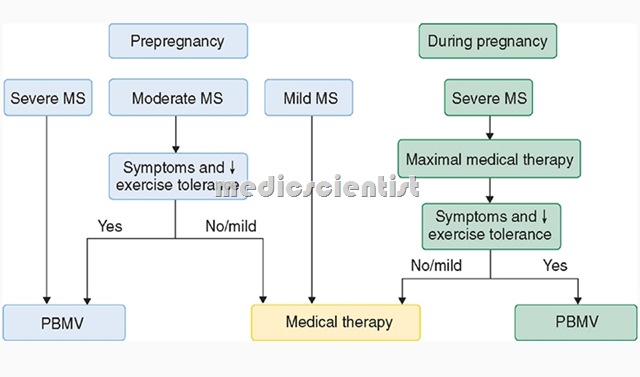
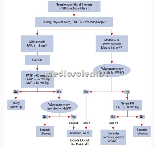
- The interventional and surgical treatment of MS is mitral valvotomy
Mitral valvotomy Indications due to Mitral Stenosis
- – Symptomatic patients
- -Mitral valve orifice less than 1 sq cm / sq m.
- body surface area.
Mitral valvotomy can be done by :
- 1. Percutaneous balloon mitral valvotomy (BMV)
- 2.Surgical valvotomy (may be closed or open) – BMV is done by passing a catheter from a leg
- vein to RA and then into LA, after transseptal puncture and a balloon put across the mitral valve and inflated to increase the valve area.
- – BMV or closed valvotomy cannot be done in patients with – adhesion of cusps, fibrosis, calcification of valve, immobile leaflets, thrombus in LA, patients with MR.
- In these patients open valvotomy is done.
- – Open valvotomy using cardiopulmonary bypass is done in patients where contraindications to BMV or closed surgical valvotomy is present.
- Mitral valve replacement: is done where valve is severely diseased and MR is present.

